An anatomical look inside brain ’P-0255’
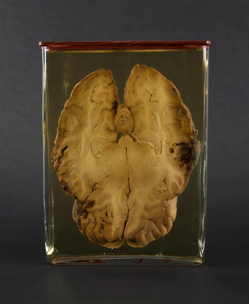
What you see in the heading image is an anatomical preparation which shows a horizontal section through the human brain with the specimen number P-0255 of the University Museum Groningen. Dark discoloration is seen on two sides: hemorrhages, probably caused by traumatic brain injury (TBI). TBI can occur when the brain collides with the skull as a result of a severe blow to the head, which can cause brain damage. This person may have fallen, had a traffic or sports accident, attempted suicide, or been assaulted.
This brain sample reveals a ’coup contre-coup’, recognizable by the two dark spots on two opposite sides.
In a blow to the head (in French: coup), the skull is accelerated against the brain so that it strikes the skull at the point of the blow. In a “contre-coup”, the brain is in motion (e.g. when falling), which is suddenly slowed down by the skull, e.g. because it hits a solid object such as a wall or the floor. In the process, the brain continues to move due to its inertia and then collides against the skull on the side opposite to the impact.
In a stronger blow, both can occur together, the brain is injured first on the side of the blow and then on the opposite site ‘coup contre-coup’, a devestating combination.
What do we know more about the brain P-0255?
This anatomical specimen is part of the private collection of the medical scientist Pieter de Riemer (1769 – 1831). De Riemer was a Dutch professor of anatomy, surgery and obstetrics, a member of the army medical board, and even the king’s ‘consultant surgeon’. During his career he built up a large collection of over 900 anatomical and physiological preparations, which were eventually donated to the University of Groningen in 1831. Back in 1818, De Riemer was the first to use the so-called Frozen Section Technique for the diagnosis of diseased tissue. With this technique, a sample can be frozen very quickly and then cut for analysis, which makes it possible, for example, to examine tissue during a surgery, and guide e.g. a tumour resection.
However, we do not know if de Riemer used this technique to assess this particular specimen, and neither what exactly happened to brain P-0255, although it would be very interesting from a neuropsychological and forensic point of view. Perhaps it was a soldier who fell to the ground in a duel on horseback and sustained a ‘coup contre-coup’ injury. He may have even came to De Riemer as a patient with a series of persistent physical and cognitive symptoms and perhaps succumbed to his TBI a short time later, which may have prompted de Riemer to examine the brain more closely to find a link to the symptoms.
The current causes of severe TBI in the Netherlands are investigated by the epidemiological BRAIN-PROJECT, a Dutch multicenter research project to improve prehospital trauma care. This is an important endeavour, as these patients are at risk of even secondary brain damage at the time before they even arrive at the hospital.
And what do you think is the main cause of severe TBI in the Netherlands? According to this project, it is neither assaults nor falls, but road traffic accidents, with bicycle accidents accounting for the largest share (Bossers et al., 2021). Fortunately, there is a fairly simple primary prevention for this: wearing bicycle helmets.
But back to P-0255. This preparation is not currently gathering dust on a shelf in storage. It is currently being examined by undergraduate psychology students in practicals as part of the Introduction to Clinical Neuropsychology course. For those interested, similar specimens can be seen in the university’s museum.
References:
Bossers, S. M., Boer, C., Bloemers, F. W., Van Lieshout, E. M., Den Hartog, D., Hoogerwerf, N., … & BRAIN-PROTECT Collaborators. (2021). Epidemiology, prehospital characteristics and outcomes of severe traumatic brain injury in the Netherlands: the BRAIN-PROTECT study. Prehospital emergency care, 25(5), 644-655.
Le Grand, J. (2022). Anatomisch Museum. Rijksuniversiteit Groningen. https://www.rug.nl/museum/collections/medical-sciences/anatomy-and-pathology
Featured image taken by Ciska Ackermann


